


01/27/2025
Electrocardiography (ECG) is one of the most essential diagnostic tools for assessing the heart's health. It records the electrical activity of the heart, which can reveal underlying conditions that may go unnoticed otherwise. While a normal ECG provides an image of a healthy heart, it can also identify abnormalities that may signal potential heart issues. When it comes to diagnosing a heart attack, the ECG test report can be a vital tool for doctors, offering clues about the extent and location of damage to the heart.
A heart attack ECG report shows distinct changes compared to a normal ECG report. Understanding the differences between these two can help you better grasp how doctors interpret your ECG and make decisions regarding treatment. Identifying the key differences between a heart attack ECG and a normal ECG, along with the significance of normal ECG values, plays a crucial role in accurate diagnosis and effective treatment of a heart attack.
What is an ECG?
Before delving into the differences between a normal ECG and a heart attack ECG, it's important to understand what an ECG is and how it works. The heart’s electrical impulses are responsible for its rhythm and contraction. These electrical signals can be measured by placing electrodes on the body, which then send the data to an ECG machine to be recorded on graph paper as a series of waves.
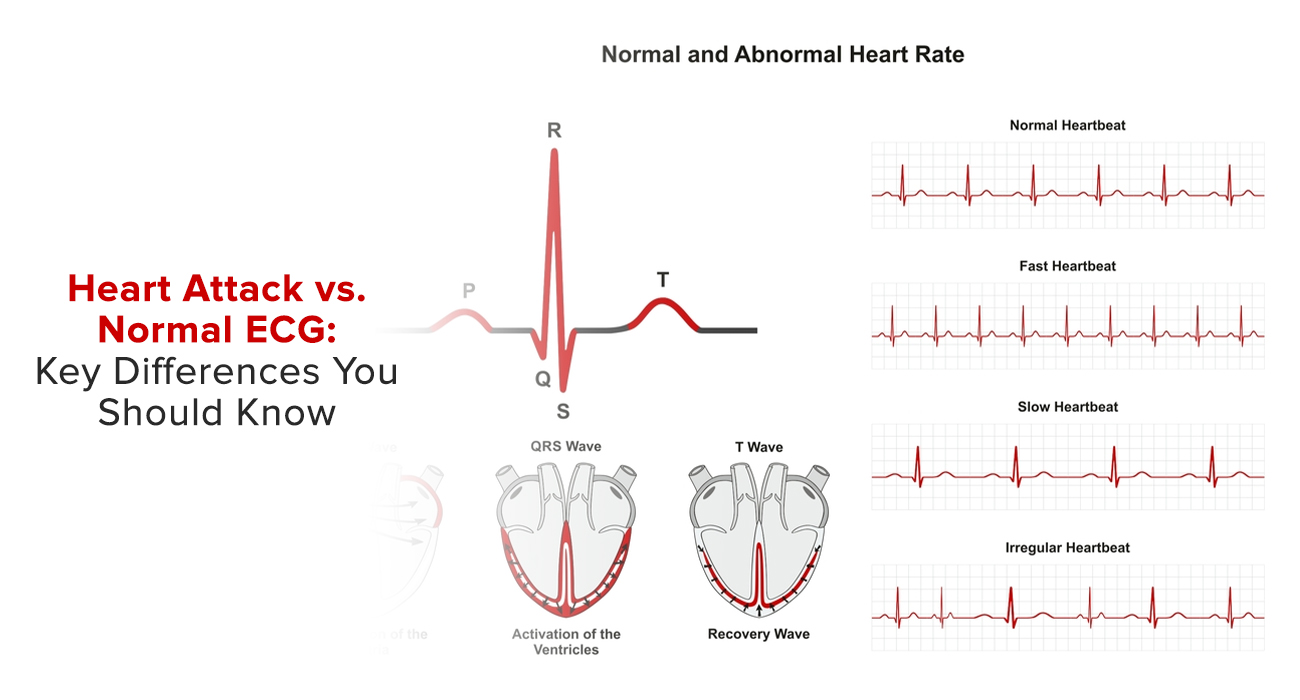
An ECG records the electrical signals from different parts of the heart and displays them on a graph, with each wave representing different phases of the heart's electrical cycle. The key components of an ECG are the P wave, QRS complex, and T wave, which correspond to the electrical activity of the heart’s atria and ventricles.
An ECG test provides doctors with important data about the heart’s rhythm, rate, and electrical activity, allowing them to detect a range of cardiac conditions. A normal ECG indicates that the heart is functioning well, while abnormal results can point to issues like arrhythmias, heart disease, or a heart attack.
What are the Key Components of a Normal ECG?
A normal ECG test report reveals a regular heart rhythm, with consistent intervals between beats. It is composed of the following elements:
- P Wave: This represents the depolarization (electrical activation) of the atria, the heart’s upper chambers.
- QRS Complex: This corresponds to the depolarization of the ventricles, which are the heart’s lower chambers.
- T Wave: The T wave reflects the repolarization or recovery of the ventricles after they contract.
In a normal ECG, the intervals between these waves are consistent and within normal limits. Typical normal ECG values include:
- Heart rate: 60-100 beats per minute (bpm)
- PR interval: 120-200 milliseconds
- QRS duration: 60-100 milliseconds
- QT interval: 350-440 milliseconds
These values are indicative of a well-functioning heart, and if your ECG falls within these ranges, it generally means your heart’s electrical system is working as it should.
What Happens During a Heart Attack?
A heart attack (myocardial infarction) occurs when blood flow to the heart muscle is blocked, typically by a blood clot or plaque buildup in one of the coronary arteries. This blockage deprives part of the heart muscle of oxygen, leading to damage or death of the heart tissue. When this occurs, the electrical activity of the heart becomes disrupted, causing notable changes in the ECG. The heart attack ECG reveals these disturbances, often presenting with patterns and abnormalities that are not seen in a normal ECG.
Key Differences Between a Normal ECG and a Heart Attack ECG
Understanding the differences between a heart attack ECG and a normal ECG is crucial in diagnosing a heart attack. The key differences are as follows:
|
Key Feature |
Normal ECG | Heart Attack ECG |
| ST Segment Elevation | Horizontal or slightly downward sloping | Elevated ST segment (more than 1 mm above baseline), indicating STEMI |
| T Wave Inversion | Upright T wave reflecting normal ventricular repolarization | Inverted T wave indicating ischemia or lack of oxygen to the heart muscle |
| Q Wave Formation | Absent or very small Q waves with no clinical significance | Pathological Q waves, larger than normal, indicating irreversible heart muscle damage (infarction) |
| Arrhythmias | Regular rhythm, consistent intervals between P, QRS, and T waves; heart rate 60–100 bpm | Irregular rhythm; arrhythmias such as ventricular tachycardia (VT) or ventricular fibrillation (VF) may be present |
| QT Interval | Within normal range of 350–440 milliseconds | Prolonged QT interval, increasing the risk of life-threatening arrhythmias |
Why ECG is Crucial in Diagnosing a Heart Attack
ECGs are an essential part of emergency care for patients suspected of having a heart attack. If a heart attack ECG report shows abnormal patterns such as ST segment elevation, T wave inversion, and pathological Q waves, it provides valuable information about the location and severity of the heart attack. In addition to the ECG, doctors may use other diagnostic tools, such as blood tests and imaging, to confirm the diagnosis.
In cases where STEMI is suspected based on the ECG test, immediate medical intervention is required to restore blood flow to the affected part of the heart. This might include procedures like angioplasty, the use of thrombolytic drugs to dissolve clots, or in some cases, surgery.
Conclusion
The differences between a heart attack ECG report and a normal ECG are significant and can provide doctors with vital information about the heart's condition. While a normal ECG shows a consistent rhythm and normal electrical activity, a heart attack ECG reveals abnormalities such as ST segment elevation, T wave inversion, Q wave formation, and arrhythmias. Recognizing these key differences allows for a quicker and more accurate diagnosis, which can be life-saving.
If you suspect you or someone you know may be experiencing a heart attack, an ECG test is one of the first steps in diagnosis. Understanding how to interpret the differences between a normal ECG and a heart attack ECG can help you better understand the importance of this tool in managing heart health. Early detection through ECG can lead to timely interventions and significantly improve outcomes for heart attack patients.
FAQs:
Q1: What is an ECG, and how does it help in detecting a heart attack?
A: An ECG (electrocardiogram) measures the heart's electrical activity and helps identify abnormalities like ST segment elevation, T wave inversion, and arrhythmias, which are key indicators of a heart attack.
Q2: Can a normal ECG rule out a heart attack?
A: Not always. While a normal ECG indicates no immediate electrical abnormalities, it cannot rule out all types of heart attacks, such as non-ST elevation myocardial infarction (NSTEMI). Further tests like blood markers may be needed.
Q3: What are the key differences between a normal ECG and a heart attack ECG?
A: A normal ECG shows consistent rhythm, normal ST segment, upright T waves, and no pathological Q waves. A heart attack ECG, however, may display ST segment elevation, T wave inversion, abnormal Q waves, and arrhythmias.
Q4: What does ST segment elevation indicate?
A: ST segment elevation is a hallmark sign of STEMI (ST-elevation myocardial infarction), a severe type of heart attack caused by complete blockage of a coronary artery.
Q5: Are all heart attacks detectable on an ECG?
A: No. While many heart attacks show distinct changes on an ECG, smaller or earlier heart attacks may not. Additional diagnostic tools, like echocardiograms or blood tests (troponin levels), are often used to confirm the diagnosis.
Q6: What should I do if my ECG shows abnormal results?
A: Consult a cardiologist immediately. Abnormal ECG findings may indicate heart conditions like arrhythmias, ischemia, or a heart attack, all of which require prompt medical attention.
Q7: How often should I get an ECG?
A: For healthy individuals, an ECG is recommended during routine check-ups if risk factors are present (e.g., hypertension, diabetes, or family history of heart disease). For those with heart conditions or symptoms, a cardiologist may advise more frequent monitoring.
Q8: Is ECG painful or risky?
A: No, an ECG is a non-invasive, painless procedure with no risks. It simply records electrical activity through electrodes placed on the skin.