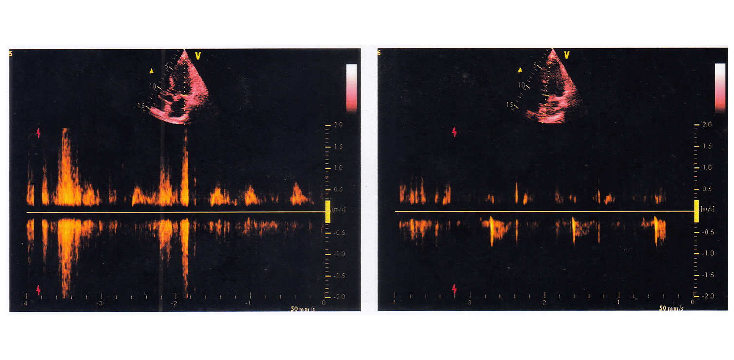
-2D Echo
A 2D echocardiogram, also known as a 2D echo, is a non-invasive diagnostic that analyzes and evaluates the function of your heart's various components. This test uses sound vibrations to produce images of various sections of the heart. It aids in determining damage, obstructions, and blood flow rates. Doctors require regular 2D echo testing to detect and treat any heart concerns early on, keeping you healthy and active as you age.
Indications
2D echocardiography is used to detect the following heart conditions:
- Any underlying heart conditions or anomalies
- Congenital cardiac disorders, blood clots, or malignancies.
- A malfunctioning heart valve
- Abnormal blood flow within the heart.
A 2D echo provides information on the operation of your heart, detects problems, and plans treatment for the growing disease.
Procedure
- The procedure takes only 30 minutes to an hour, is completely safe, and is carried out under the supervision of a radiologist and cardiologist.
- The technician in the lab applies a colorless gel and moves the transducer across the various areas of the chest to evaluate the sound vibrations using a transducer.
- The gel enables the transducer to examine the heart, its tissues, and its structures.
- Following the test, the gel is thoroughly cleansed.
- The test images will be either printed on paper or recorded on DVD. As a result, your doctor can evaluate the findings for abnormalities or signs of disease.


 0141-3120000